Uncommon Thoracic HRCT Pattern in a Patient with Anti-MDA5 Myositis-Related Interstitial Lung Disease
by Anastasios Palamidas*, Demosthenes Bouros
Department of Pneumonology, Athens Medical Center and Medical School, University of Athens, Greece
*Corresponding Author: Anastasios Palamidas, Department of Pneumonology, Athens Medical Center and Medical School, University of Athens, Greece
Received Date: 11 February 2026
Accepted Date: 16 February 2026
Published Date: 18 February 2026
Citation: Palamidas A, Bouros D. (2026). Uncommon Thoracic HRCT Pattern in a Patient with Anti-MDA5 Myositis-Related Interstitial Lung Disease. Ann Case Report. 11: 2538. DOI: https://doi.org/10.29011/2574-7754.102538
Introduction
A 58-year-old with a history of nonspecific interstitial pneumonia (NSIP) pattern in high-resolution computed tomography (HRCT) (Figure 1A) presented with dyspnea and hypoxemia (SaO2 85% rest). The patient was initially followed up in another hospital; his routine serology was negative, and received treatment with mycophenolate mofetil in 2017 based on HRCT findings. Physical examination revealed bibasilar “Velcro” crackles and a V-shaped shawl erythematous rash over the chest. Pulmonary function testing demonstrated a significant decline from 2017 to 2025 in forced vital capacity (FVC: 63%pred to 53%pred) and diffusing capacity (Dlco: 55%pred to 34%pred).
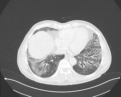
Figure 1A: CT SCAN (2017): NSIP pattern bilaterally at the lung bases depicting traction bronchiectasis, fine reticulation, ground glass opacities, with subpleural sparing.
A new HRCT revealed a strikingly asymmetric fibrotic pattern a fibrotic NSIP involving the right lung, in contrast to an exuberant usual interstitial pneumonia (UIP) pattern in the left lung (Figure 1B). Given the cutaneous findings, the deteriorating clinical course and the uncommon radiologic findings, extended serologic testing, including a myositis panel, was repeated, demonstrating a positivity for anti–MDA5 antibodies.
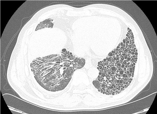
Figure 1B: HRCT scan (2025): A striking atypical asymmetric pattern of a fibrotic NSIP at the right lung base and an exuberant UIP pattern at the left lung.
Anti-MDA5 myositis is associated with dermatomyositis and progressive interstitial lung disease, characterized by high early mortality, poor response to immunosuppression, and minimal (amyopathic) muscle involvement [1]. Radiologically, organizing pneumonia and NSIP predominate, whereas UIP is uncommon [2], rendering the unilateral UIP pattern in this case particularly notable. Antifibrotic therapy (nintedanib) was added to ongoing MMF treatment.
References
- Lu X, Peng Q, Wang G. (2024). Anti-MDA5 antibody-positive dermatomyositis: pathogenesis and clinical progress. Nat Rev Rheumatol. 20: 48-62.
- Chen X, Jiang W, Jin Q, Peng Q, Zhang L, et al. (2023). Clinical, radiological, and pathological features of anti-MDA5 antibody-associated interstitial lung disease. RMD Open. 9: e003150.
© by the Authors & Gavin Publishers. This is an Open Access Journal Article Published Under Attribution-Share Alike CC BY-SA: Creative Commons Attribution-Share Alike 4.0 International License. Read More About Open Access Policy.