Superimposing Intraoral and Facial Digital Scan: A Novel Technique for Accurate Spatial Alignment of Dental Arches
by Marco Valenti1*, Alessandro Valenti, Laura Canale2, Johannes H Schmitz3, Davide Cortellini4
1Private practice in Pordenone, via GB Damiani 5, Italy
2Dental Lab owner in Rimini, via Luigi Tonini 18, Italy
3Private practice in Milan, Galleria Buenos Aires 14, Italy
4Private practice in Riccione, Via Ippolito Nievo 11, Italy
*Corresponding author: Marco Valenti, Private practice in Pordenone, via GB Damiani 5, Italy
Received Date: 26 March, 2024
Accepted Date: 01 April, 2024
Published Date: 03 April, 2024
Citation: Valenti M, Valenti A, Canale L, Schmitz JH, Cortellini D, et al. (2024) Superimposing Intraoral and Facial Digital Scan: A Novel Technique for Accurate Spatial Alignment of Dental Arches. Dent Adv Res 9: 100208. https://doi.org/10.29011/2574-7347.100208
Abstract
A reliable novel technique for superimposing intraoral and face digital scan without using any aligner or scan abutment is presented. This technique involves scanning the perioral area by incorporating parts of the upper dental arch into the perioral tissue scan. Unlike other proposed methods, including the upper dental arch scan in the perioral tissues scan, ensure accurate spatial positioning of intraoral dental arches scans within the facial scan.
Keywords: Alignment point tool, Facial digital scan, Face scanner, Intraoral scan, Superimposing
Introduction
Assessing facial and dental structures is crucial for achieving successful esthetic prosthodontic treatments. A Two-Dimensional (2D) esthetic analysis utilizing facial and dental photographs has been introduced to ensure predictable esthetic outcomes [1-4]. This analysis involves importing full-face photographs taken in a relaxed state, during a wide smile, and smartphone video editing software that supports the smile design for an esthetic treatment plan [5].
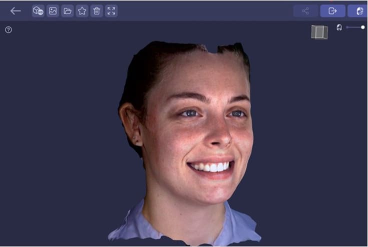
Advancements in Three-Dimensional (3D) facial scanning have made it possible to virtually plan a prosthetic design that aligns with the corresponding facial appearance in a 3-D perspective [6]. Through 3D facial scanning, a virtual face can be generated and seamlessly integrated with 3D dental images obtained from digital scans of the teeth [7].
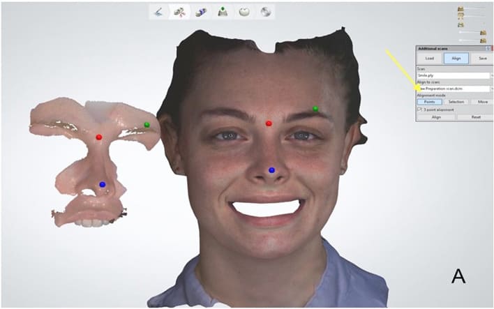
Many different techniques were proposed to superimpose the intraoral scan into a facial scan to create a 3-dimensional virtual patient, using aligner systems or scan abutment [8-10] or simply collecting additional facial scans and perioral scans to collect some skin and dental references merging in superimposition [11,12].
This article presents a novel technique for superimposing facial and intraoral scans, eliminating the need for aligners, scan bodies, or extraoral landmarks. It works well even in patients who do not expose upper incisors smiling. Furthermore, this technique enables precise spatial alignment of intraoral scans within facial scans.
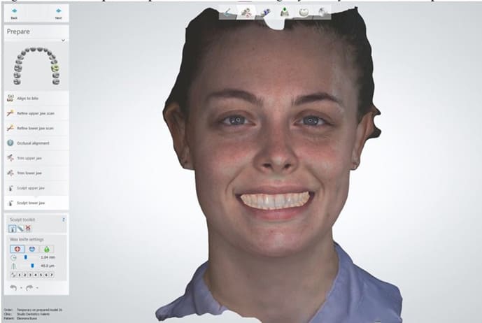
Technique
To simplify the digitalization and superimposing of the facial and intraoral scans to obtain a 3D virtual representation of a patient, the subsequent protocol was performed:
Discussion
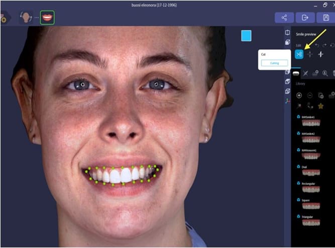
In prosthetic treatments involving the esthetic area, the challenge for the dental technician and dentist is to precisely define the interpupillary and midsagittal planes to correctly establish ideal teeth shape concerning the patient’s smile line and lips position (Figure 10). Facial scanning, in comparison to the 2D photography, offers greater accuracy [14], and by incorporating 3D restorations design onto the 3D facial file, it is possible to assess the shapes of the teeth on the x-y-z axis and evaluate their aesthetic outcome before testing it on the patient with interim restorations or resin mock-up. Both facial scanning and its superimposition with intraoral scanning must be accurate to perform these assessments [15].
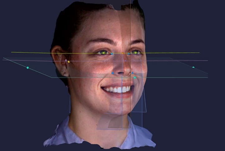
Figure 10: Positioning inter pupillary, midsagittal, and Camper planes on the facial scan helps the dentist and technician in determining the ideal teeth shape in relation to the patient’s smile line and lips position.
Conversely to a smartphone-specific application that uses a 3D sensor camera [10,12,16] or industrial scanners adapted for medical use [17,18] the advantage of using a static face scanner (Rayface 100,200; Ray Co., Ltd) is that the patient remains still and in 0,5 seconds the scanner scans patient’s face, reducing the risk of inaccurate scans due to movements by the patient, scanner or operator [19,20].
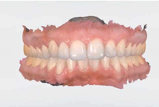
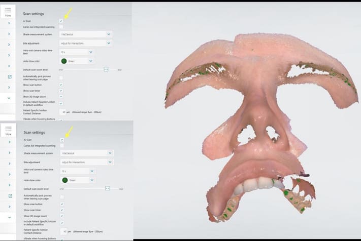
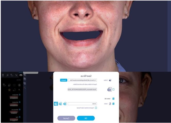
An accurate facial scan [21] is not enough because, due to the distortion all face scanners introduce in the patient’s teeth, it is impossible to superimpose a facial scan to the patient’s dental arches scan. The key to this technique’s success is using a perioral scan that includes lips, nose, forehead, and teeth. The accuracy and time scan of the IOS (TRIOS 3, 4, or 5; 3Shape A/S) [22,23] allow for simple and reliable perioral scanning, avoiding any involuntary patient movements that would affect the accuracy of the perioral scan.
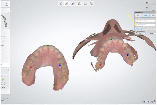
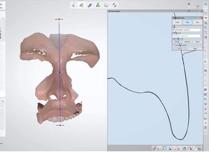
Compared to the technique proposed by Lo Russo, et al. [12] scanning the posterior teeth also allows for the correct sagittal and horizontal alignment of the arches during the superimposition with the facial scan. Furthermore, thanks to the CAD-CAM software program (Dental System; 3 shape A/S) cross-sectional tool, it is possible to assess the accuracy of alignment and correct it by slightly shifting any of the 3 superimposed scans, reducing the risk of mismatching.
Creating a 3-D virtual patient representation could help dentists and dental technicians improve diagnosis, facially driven treatment plans, and prosthetic treatment outcomes. Even if most of the scanners available on the market exhibit accuracy values that are acceptable for clinical application, [24] the 3-D virtual patient accuracy and trueness could be influenced by techniques used and operator skill [25]. The proposed technique simplifies the superimposing procedures to align intraoral scans with facial scans, making the creation of the virtual patient repeatable and reliable without using skin landmarks or scan abutments.
Summary
A technique that allows clinicians and dental technicians to obtain valuable information regarding the relationship between patient smile and patient face is presented. Intraoral, perioral, and facial scans are superimposed easily without any scan abutment, aligner, or skin landmarks, with a CAD software program and an alignment point tool. Furthermore, the spatial alignment of dental arches on facial scan could be more precise than other techniques proposed, thanks to the perioral scan that should include the upper premolar and molar teeth surface and not only incisors.
Declaration
Patient consent
Written informed consent was obtained prior to capturing any face scan or images and published.
Conflict of interest
Authors declare no conflict of interest.
References
- Nold SL, Horvath SD, Stampf S, et al. (2014) Analysis of select facial and dental esthetic parameters. Int J Periodontics Restorative Dent 34: 623-629.
- Milutinovic J, Aleksic E, Avramov S, et al. (2023) Esthetic preferences of orthodontists, dentists, and plastic surgeons for balanced facial profiles. J Oral Sci 65: 73-76.
- Koidou VP, Chatzopoulos GS, Rosenstiel SF, et al. (2018) Quantification of facial and smile esthetics. J Prosthet Dent 119: 270-277.
- Frese C, Staehle HJ, Wolff D (2012) The assessment of dentofacial esthetics in restorative dentistry: A review of the literature. J Am Dent Assoc 143: 461-466.
- Coachman C, Calamita MA, Sesma N (2017) Dynamic documentation of the smile and the 2D/3D digital smile design process. Int J Periodontics Restorative Dent 37: 183-193.
- Sanchez-Lara A, Chochlidakis KM, Lampraki E, et al. (2019) Comprehensive digital approach with the Digital Smile System: A clinical report. J Prosthet Dent 121: 871-875.
- Yuan Y, Liu Q, Yang S, et al. (2023) Four-dimensional superimposition techniques to compose dental dynamic virtual patients: A systematic review. J Funct Biomater 6: 14:33
- Amezua X, Erkizia G, Jauregi M, et al. (2022) Creating threedimensional virtual patients by superimposing intraoral and facial digital scans guided with an aligner system: A dental technique. J Prosthet Dent 31: 00611-614.
- Perez-Giugovaz MG, Park SH, Revilla-Leon M. (2021) Threedimensional virtual representation by superimposing facial and intraoral digital scans with an additively manufactured intraoral scan body. J Prosthet Dent 126: 459-463.
- Revilla-Leon M, Zandinejad A, Nair MK, et al. (2022) Accuracy of a patient 3-dimensional virtual representation obtained from the superimposition of facial and intraoral scans guided by extraoral and intraoral scan body systems. J Prosthet Dent 128: 984-993.
- Hassan B, Gimenez Gonzalez B, Tahmaseb A, et al. (2017) A digital approach integrating facial scanning in a CAD-CAM workflow for complete-mouth implant-supported rehabilitation of patients with edentulism: A pilot clinical study. J Prosthet Dent 117: 486-492.
- Lo Russo L, Di Gioia C, Salamini A, et al. (2020) Integrating intraoral, perioral, and facial scans into the design of digital dentures. J Prosthet Dent 123: 584-588.
- Valenti M, Schmitz JH (2021) A reverse digital workflow by using an interim restoration scan and patient-specific motion with an intraoral scanner. J Prosthet Dent 126: 19-23.
- Cascos R, Ortiz Del Amo L, Álvarez-Guzmán F, et al. (2023) Accuracy between 2D photography and dual-structured light 3D facial scanner for facial anthropometry: A Clinical Study. J Clin Med 12: 3090.
- Amezua X, Iturrate M, Garikano X, et al. (2023) Analysis of the impact of the facial scanning method on the precision of a virtual facebow record technique: An in vivo study. J Prosthet Dent 130: 382-391.
- Pellitteri F, Brucculeri L, Spedicato GA, et al. (2021) Comparison of the accuracy of digital face scans obtained by two different scanners. Angle Orthod 91: 641-649.
- Otawa N, Aoki T, Sumida T, et al. (2022) Application of a new scan body for face-driven fixed prosthetics. Clin Exp Dent Res 8: 275-281.
- Michelinakis G, Apostolakis D, Velidakis E (2023) An in vitro comparison of accuracy between three different face scanning modalities. Eur J Prosthodont Restor Dent 15.
- Mai HN, Kim J, Choi YH, et al. (2020) Accuracy of portable facescanning devices for obtaining three-dimensional face models: A systematic review and meta-analysis. Int J Environ Res Public Health 18: 94.
- Franco de Sa Gomes C, Libdy MR, et al. (2019) Scan time, reliability and accuracy of craniofacial measurements using a 3D light scanner. J Oral Biol Craniofac Res 9: 331-335.
- Cho RY, Byun SH, Yi SM, et al. (2023) Comparative analysis of three facial scanners for creating digital twins by focusing on the difference in scanning method. Bioengineering (Basel) 10: 545.
- Roth I, Hermann P, Vitai V, et al. (2023) Comparison of the learning curve of intraoral scanning with two different intraoral scanners based on scanning time. BMC Oral Health 23: 267.
- Thomas AA, Jain RK. Influence of operator experience on scanning time and accuracy with two Different intraoral scanners: A prospective clinical trial. Turk J Orthod 36: 10-14.
- Bohner L, Gamba DD, Hanisch M, et al. (2019) Accuracy of digital technologies for scanning facial, skeletal, and intraoral tissues: A systematic review. J Prosthet Dent 121: 246-251.
- Revilla-Leon M, Zeitler JM, Barmak AB, et al. (2022) Accuracy of the 3-dimensional virtual patient representation obtained by using 4 different techniques: An in vitro study. J Prosthet Dent S0022-3913: 00342.
© by the Authors & Gavin Publishers. This is an Open Access Journal Article Published Under Attribution-Share Alike CC BY-SA: Creative Commons Attribution-Share Alike 4.0 International License. Read More About Open Access Policy.