Spontaneous Rupture of the Extensor Pollicis Longus Tendon Caused by a Wrist Sprain
by Jin Young Kim*, Min Sung Kum
Department of Orthopaedic Surgery, Dongguk University College of Medicine, Korea
*Corresponding author: Jin Young Kim, Professor of Orthopaedic Surgery, Dongguk University Ilsan hospital Ilsan, Goyang, Korea
Received Date: 19 November, 2024
Accepted Date: 02 December, 2024
Published Date: 05 December, 2024
Citation: Kim JY, Kum MS (2024) Spontaneous Rupture of the Extensor Pollicis Longus Tendon Caused by a Wrist Sprain. J Orthop Res Ther 9: 1369. https://doi.org/10.29011/2575-8241.001369
Abstract
The aim of this case report is to show an extensor pollicis long rupture caused by a wrist sprain. The patient sustained wrist dorsiflexion injury while riding a bicycle and complained of inability of thumb extension 5 weeks after the injury. There was no fracture in distal radius on plain radiographs and MRI. However, we found EPL rupture around the Lister’s tubercle, which was treated by EIP transfer. We are carefully suggesting that a wrist can result in a spontaneous rupture of EPL, if it is severe enough to accompany significant hematoma or edema around the EPL, even though distal radius fracture does not exist.
Keywords: Extensor pollicis longus; Spontaneous rupture; Sprain; Wrist Sprain; Tendon transfer.
Introduction
Some authors mentioned that spontaneous or delayed rupture of the extensor pollicis longus (EPL) tendon is not uncommon and is associated with rheumatoid arthritis, fractures of the wrist, systemic or local steroid injections and repetitive and excessive abnormal motion of the wrist joint [1,2,3]. However, EPL rupture caused by a minor trauma such as a wrist sprain is quite rare, especially without predisposing factor such as a non-displaced distal radius fracture. We experienced a patient who was presented with an EPL rupture 5 weeks after a wrist sprain. Thus, we report this case with a review of the previous literatures.
Case Presentation
A 36-year-old male patient visited our clinic, complaining of right wrist pain. He was an office worker and had no medical history on his right wrist. He rode his bicycle and bumped into a car which was parked in a street one day before the visit. The handle of the bicycle hit his palm quite hard making his wrist hyperextended. Physical examination revealed moderate tenderness and swelling on the dorsal aspect of the wrist. Active motion, especially dorsiflexion was limited due to pain. However, the plain radiographs at initial visit showed no definite fracture at his right wrist but soft tissue swelling of the wrist dorsum (Figure 1). We diagnosed it as a wrist sprain and put a short arm splint on his right wrist. We checked plain radiographs again 2 weeks after the injury. As we confirmed that there was no fracture identified on the plain radiographs, we removed the splint and let him return to his daily activity. However, he visited our clinic 5 weeks after injury, complaining of inability to actively extend his right thumb. Physical examination showed loss of active extension of thumb interphalangeal joint. On MRI, an EPL tendon was ruptured around the Lister’s tubercle (Figure 2), although there was no signal change of distal radius or other carpal bones, suggesting a bone contusion or a microfracture. The distal stump was identified around the 1st carpo-metacarpal joint, and the proximal stump was not definitely delineated. Abundant fluid collection was observed around the ruptured tendon ends (Figure 2).
Surgery was performed under regional (brachial plexus block) anesthesia. An incision was made over the 3rd compartment just ulnar to Lister’s tubercle. The retinaculum of 3rd compartment was divided longitudinally. We found the proximal stump of ruptured EPL tendon slightly proximal to the Lister’s tubercle. The proximal stump was 3cm long and very thin, which had little tendon tissue and was surrounded with thick synovial sheath (Figure 3). Second incision was made over the snuff box. The distal stump of ruptured EPL tendon was found at the dorsum of radio-scaphoid joint. The torn tendon ends were quite thinned and fraying (Figure 3), and end-to-end anastomosis of the injured tendon was not feasible. We performed tendon transfer surgery using extensor indicis proprius (EIP) tendon. Postoperative short arm thumb-spica splint was applied for 4 weeks with the wrist in neutral and the thumb extended and abducted. The thumb was gradually permitted to move with a removable splint for 4 weeks.
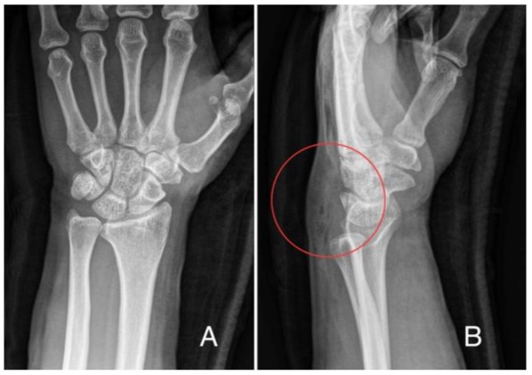
Figure 1: The plain radiographs at initial visit showed no definite fracture at both wrists (A) Postero-anterior (B) Lateral (red circle): Soft tissue swelling of the wrist dorsum was observed.
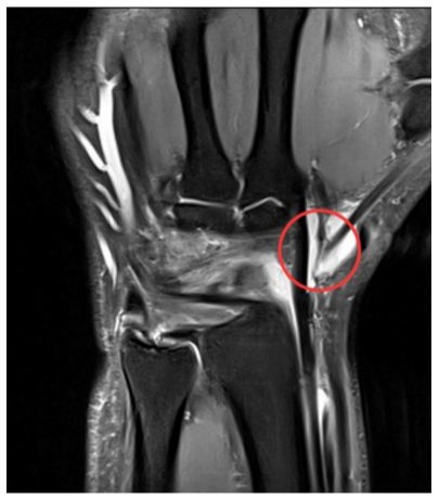
Figure 2: On MRI (T2 Coronal), an EPL tendon rupture was observed and the distal stump was identified around the 1st carpometacarpal joint (red circle). Fluid collection was observed around the ruptured tendon ends and there was no signal change of distal radius or other carpal bones, suggesting a bone contusion or a micro-fracture.
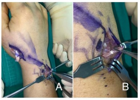
Figure 3: The torn EPL tendon ends were identified (A). The proximal stump was found slightly proximal to the Lister’s tubercle and the distal stump at the dorsum of radio-scaphoid joint. The proximal stump (white Asterix) was 3cm long and very thin, which had little tendon tissue and was surrounded with thick synovial sheath (B).
Discussion
Spontaneous rupture of the EPL tendon commonly occurs in different situations such as rheumatoid arthritis, systemic or local steroid injections, tophaceous gout infiltration, ankylosing spondylitis, and misplaced external fixators or metal plates [1,3,4]. It is also well known that a non-displaced distal radius fracture was the most common cause of the spontaneous rupture of the EPL tendon in trauma-related cases. However, a rupture of the EPL tendon caused by wrist dorsiflexion injury, such as a sprain without distal radius fracture, was quite rarely reported. Bjorkman and Jorgsholm [1] reported three cases of EPL rupture by blunt trauma without distal radius fracture. However, one of them hit his wrist directly on a light arc and two patients had stab wound on the wrists caused by sharp objects. Ching-Hsuan et al. [5] also reported four EPL ruptures having a history of falling onto an outstretched wrist. However, they mentioned that they couldn’t exclude an occult distal radius fracture using CT scan, even though a prominent Lister tubercle was not found on the plain radiographs.
There are two major mechanisms explaining the pathogenesis of spontaneous rupture of the EPL tendon. One is mechanical theory and the other is vascular theory [1,6]. According to the mechanical theory, the EPL tendon courses over the dorsal cortex around Lister’s tubercle and the rough bony edge at the distal radius fracture abrades the EPL tendon [1,7]. The vascular theory says that the increased pressure within the tendon sheath at Lister’s tubercle causes ischemia and delayed rupture of the EPL tendon [1,6,7]. In our patient, we could not find a definite fracture of distal radius or carpal bones not only on x-ray but also on MRI. MRI showed that EPL tendon rupture around the Lister’s tubercle and abundant fluid collection around the torn tendon ends. Thus, we presumed that significant hematoma caused by a dorsiflexion injury of a wrist led to subsequent pressure increase on the EPL tendon, which resulted in EPL the tendon rupture.
Conclusion
We think that this is another exemplary case supporting that blunt trauma (dorsiflexion injury) of the wrist could cause rupture of EPL tendon without distal radius fracture. Based on MRI finding, we are carefully suggesting that a wrist sprain by forced hyperextension can result in delayed EPL tendon rupture if it accompanies significant swelling, even though distal radius fracture does not exist.
Declaration
Contributors
All authors contributed to planning, reviewing and writing this article. All authors agreed to submit this article.
Conflict of interests
The authors declare no potential conflicts of interest with respect to the research, authorship, and/or publication of this article. Conflict of interest: None declared.
Ethical approval
Not applicable
Funding
This research received no specific grant from any funding agency in the public, commercial, or not-for-profit sectors.
References
- Bjorkman A, Jorgsholm P (2004) Rupture of the extensor pollicis longus tendon: a study of aetiological factors. Scand J Plast Reconstr Surg Hand Surg 38(1): 32-35.
- Harvey FJ, Harvey PM (1989) Three rare causes of extensor tendon rupture. J Hand Surg Am 14(6): 957-962.
- Taş S, Balta S, Benlier E (2014) Spontaneous rupture of the extensor pollicis longus tendon due to unusual etiology. Balkan Med J 31(1): 105-106.
- Hung JY, Wang SJ, Wu SS (2005) Spontaneous rupture of extensor pollicis longus tendon with tophaceous gout infiltration. Arch Orthop Trauma Surg 125(4):281-284.
- Ching-Hsuan Hu, Fufa D, Hsu CC, Lin YT, Lin CH (2015) Revisiting spontaneous rupture of the extensor pollicis longus tendon: eight cases without identifiable predisposing factor. Hand (N Y) 10(4): 726731.
- Choi JC, Kim WS, Na HY, Lee YS, Song WS, et al. (2011) Spontaneous rupture of the extensor pollicis longus tendon in a tailor. Clin Orthop Surg 3(2): 167-169.
- Engkvist O, Lundborg G (1987) Rupture of the extensor pollicis longus tendon after fracture of the lower end of the radius: a clinical and microangiographic study. Hand 11(1): 76-86.
© by the Authors & Gavin Publishers. This is an Open Access Journal Article Published Under Attribution-Share Alike CC BY-SA: Creative Commons Attribution-Share Alike 4.0 International License. Read More About Open Access Policy.