Objective Evaluation of Nasal Tip Reduction Using Three-Dimensional Imaging Analysis Software; Pilot Study
by Takahiko Tamura1*, Taichi Tamura1, Kohki Okumura1, Hiroo Teranishi1
Tokyo Chuo Beauty Clinic Umeda Osaka Ekimae Clinic, Osaka, Japan
*Corresponding Author: Takahiko Tamura, Department of Tokyo Chuo Beauty Clinic Umeda Osaka Ekimae Clinic, Osaka, Japan
Received Date: 24 March 2026
Accepted Date: 30 March 2026
Published Date: 01 April 2026
Citation: Tamura T, Tamura T, Okumura K, Teranishi H (2026) Objective Evaluation of Nasal Tip Reduction Using Three-Dimensional Imaging Analysis Software; Pilot Study J Surg 11: 11597 DOI: https://doi.org/10.29011/2575-9760.011597
Abstract
Background: Nasal tip reduction is an impactful procedure in esthetic facial surgery; its demand has been annually increasing. Traditional assessment methods relying on Two-Dimensional (2D) photographic images and subjective evaluation criteria have limited spatial accuracy. This study aimed to objectively quantify the postoperative volumetric changes following nasal tip reduction using Three-Dimensional (3D) imaging analysis software.
Methods: One male and six female patients (N=7) underwent nasal tip reduction surgery at our institution between March and August 2024. Volumetric changes in the nasal tip were measured using a VECTRA H2 3D imaging system. Measurements were performed at five time points: preoperatively; immediately postoperatively; and 1–2 weeks, 1 month, and 3 months postoperatively. The primary outcome was the change in nasal tip volume from the baseline.
Results: The mean age of the participants was 24.3±3.2 years. The nasal tip volume decreased consistently over time. The volume reduction was 0.17±0.18 mL (right: 0.07±0.10; left: 0.09±0.11; n=7) immediately after the surgery; 0.12±0.06 mL (right: 0.05±0.01; left: 0.06±0.05; n=7) at 1–2 weeks; 0.12±0.06 mL (right: 0.05±0.04; left: 0.08±0.04; n=6) at 1 month; and 0.08±0.06 mL (right: 0.04±0.02; left: 0.05±0.04; n=2) at 3 months.
Conclusion: The 3D imaging analysis software enabled an accurate and objective evaluation of volumetric changes following nasal tip reduction. This noninvasive method could standardize postoperative assessments, improve surgical validation, and enhance patient satisfaction.
Keywords: 3D Imaging; Facial Surgery; Nasal Tip Reduction; Rhinoplasty
Introduction
Rhinoplasty has gained popularity in Japan in recent years and is now one of the most frequently performed aesthetic procedures [1]. According to the 2023 report by the International Society of Aesthetic Plastic Surgery (ISAPS), rhinoplasty was among the top five popular procedures worldwide in 2021 [2]. The 2024 report by the Japan Society Of Aesthetic Plastic Surgery (JSAPS) ranked rhinoplasty fourth among surgical procedures in 2023, following blepharoplasty, facelift, and fat injection, with 13,073 cases accounting for 7.4% of all surgeries [3].
Nasal tip reduction requires a detailed understanding of multilayer anatomical structures, including soft tissue, cartilage, and skin. Subtle changes in the nasal tip can significantly impact facial aesthetics. Therefore, accurate evaluation of the postoperative outcomes is essential. Conventional assessment methods primarily rely on Two-Dimensional (2D) photographs and subjective interpretations. Although indices such as Goode’s ratio and the alar-nasal length proportion are commonly used, these are limited by variability and do not provide in-depth information [4,5]. The demand for more precise and objective surgical assessment is increasing. Three-dimensional (3D) imaging analysis offers reproducible high-resolution volumetric data and has shown promise for postoperative evaluation and treatment planning [6]. In this study, we used 3D imaging analysis software to quantify changes in the nasal tip volume before and after reduction surgery with the aim to validate its accuracy.
Materials And Methods
This prospective, observational study was conducted at a single institution. Patients who underwent nasal tip reduction between March and August, 2024, were included in this study (N= 7). In all patients, 3D facial images were captured using the VECTRA H2 system (Canfield Scientific, USA); the nasal tip volumes on both sides of the face were assessed (Figure 1). Measurements were obtained at baseline (preoperative), immediately after surgery, and 1–2 weeks and 1 and 3 months postoperatively. Changes in the nasal tip volume were recorded and analyzed. Descriptive statistics were performed using SPSS version 28.0 (IBM Corp., Armonk, NY, USA). All results are presented as mean±standard deviation.
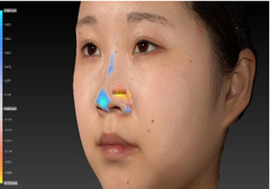
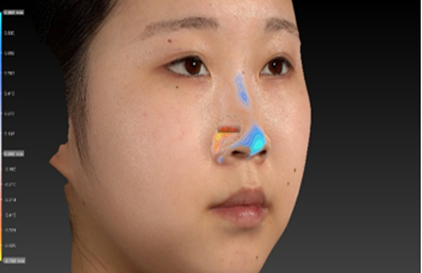
Figure 1: Nasal tip measurements using a 3D imaging system. A) Left side, B) Right side. white circle area: measurement area; blue area: volume gain area; and red area: volume loss area. 3D, three-dimensional.
Results
The study included seven patients (one male and six female participants) with a mean age of 24.3±3.2 years. The number of patients available for evaluation at each time point was: preoperative/postoperative (N=7), 1–2 weeks (n=5), 1 month (n=6), and 3 months (n=2). The mean volume reduction in the nasal tip was 0.17±0.18 mL immediately after surgery (right: 0.07±0.10 mL; left: 0.09±0.11 mL), 0.12±0.06 mL at 1–2 weeks (right: 0.05±0.01 mL; left: 0.06±0.05 mL), 0.12 ± 0.06 mL at 1 month (right: 0.05±0.04 mL; left: 0.08±0.04 mL), and 0.08±0.06 mL at 3 months (right: 0.04±0.02 mL; left: 0.05±0.04 mL).
Discussion
The clinical utility of 3D imaging technology has gained recognition in the field of aesthetic medicine. Traditional photographic evaluations are inherently subjective and limited to 2D data, which restricts the accurate analysis of spatial and volumetric changes [4,5]. This study is the first to quantify nasal tip volume reduction using 3D imaging software in a noninvasive manner. Oliveira et al. demonstrated that 0.05–0.1 mL of hyaluronic acid filler could significantly improve nasal tip projection and definition in nonsurgical rhinoplasty [7]. Our findings indicated that postoperative volume changes averaging approximately 0.12 mL are clinically meaningful and likely contribute to aesthetic improvement. The effect of volume reduction was sustained for up to 3 months in this cohort. The utility of 3D imaging has also been demonstrated in other surgical fields. Lo et al. applied machine learning to 3D facial images to assess symmetry before and after orthognathic surgery [8], whereas Zupan et al. measured soft tissue changes after maxillary expansion using 3D analysis [9]. In our study, the observed volume reduction correlated with progressive healing and decreased edema, suggesting that initial postoperative changes reflect soft tissue swelling, whereas later reductions indicated structural remodeling. The 3D imaging could provide reproducible and objective data, allowing precise visualization of surgical outcomes and facilitating patient communication (Table 1). Unlike computed tomography, this method is noninvasive and suitable for serial follow-ups. The study was limited by its small sample size and the absence of statistical testing, due to the pilot nature of the analysis. Future studies should include larger cohorts, multicenter collaborations, and integration with patient-reported outcomes.
|
Post-operation |
1,2 week |
1M |
3M |
|
|
(N=7) |
(n=5) |
(n=6) |
(n=2) |
|
|
Volume change (R) |
0.07±0.10 |
0.05±0.01 |
0.05±0.04 |
0.04±0.02 |
|
Volume change (L) |
0.09±0.11 |
0.06±0.05 |
0.08±0.04 |
0.05±0.04 |
|
Volume change (total) |
0.17±0.18 |
0.12±0.06 |
0.12±0.06 |
0.08±0.06 |
L, left; R, right; 3D, three-dimensional
Table 1: Volume changes measured using 3D imaging system (mL).
Conclusion
This is the first study to non-invasively and quantitatively evaluate volumetric changes following nasal tip reduction. The 3D imaging analysis offers a reliable, reproducible, and high-resolution method for surgical assessment and can improve the quality of postoperative care and patient satisfaction in esthetic plastic surgery.
Ethical approval: All procedures performed in studies involving human participants were in accordance with the ethical standards of the institutional and/or national research committee and with the 1964 Helsinki declaration and its later amendments or comparable ethical standards. The study protocol was approved by the institutional ethics committee (approval no.: UMEDAERB-2025Apr005).
Informed consent: All patients provided written informed consent for the surgical procedures as well as for the use of anonymized data and clinical images in academic presentations and publications.
Acknowledgments: We would like to thank Editage (www.editage.jp) for English language editing.
Conflict of interest: The authors declare that they have no conflict of interest.
Funding:None
References
- Funakoshi Y, Saito M, Kawaguchi K, Hiramatsu E, Yamamoto N, et al. (2023) Recent status of procedures in a single nationwide cosmetic surgery group. Plast Reconstr Surg Glob Open 11: e5330.
- International Society of Aesthetic Plastic Surgery (ISAPS) (2023) ISAPS International Survey on aesthetic/cosmetic procedures performed in 2021. Aesthetic Plastic Surgery. https://www.isaps.org/wp-content/uploads/2023/12/ISAPS-Global-Survey_2021.pdf.
- Japan Society of Aesthetic Plastic Surgery (JSAPS) (2024) 2024 Annual Report on Cosmetic Surgery Trends in Japan.
- Lee SH, Cho J, Lee JS (2022) Long-term outcomes of secondary nasal tip plasty after degradation of a polycaprolactone (PCL) mesh. Aesthetic Plast Surg 46: 2358-2365.
- Kılınç DD, Sayar G (2022) Evaluation of nasal tip projection and rotation of nasal tip after orthognathic surgery by using Goode's method. J Maxillofac Plast Reconstr Surg 21: 510-514.
- Okumura K, Tamura T, Funakoshi Y, Teranishi H (2024) Efficacy of fibrin sealant in submental liposuction: A prospective randomized study. Aesthetic Plast Surg
- Oliveira LL, Braz A, Palermo E, Issa MCA (2025) Nonsurgical elementary rhinoplasty: A volumetric standardized hyaluronic acid filling technique in five steps Indian J Plast Surg 58: 139-145.
- Lo LJ, Yang CT, Ho CT, Liao CH, Lin HH (2021) Automatic assessment of 3-dimensional facial soft tissue symmetry before and after orthognathic surgery using a machine learning model: A preliminary experience. Ann Plast Surg 86: S224-S228.
- Zupan J, Ihan Hren N, Verdenik M (2022) An evaluation of three-dimensional facial changes after surgically assisted rapid maxillary expansion (SARME): an observational study. BMC Oral Health 22:155.
© by the Authors & Gavin Publishers. This is an Open Access Journal Article Published Under Attribution-Share Alike CC BY-SA: Creative Commons Attribution-Share Alike 4.0 International License. Read More About Open Access Policy.