Idiopathic Mondor’s Disease of the Breast in A 35-Year-Old Woman: A Case Report and Literature Review
by Aikaterini Koufonikola, Noursen Kechagia, Sevgki Batzak, Dimitrios Karamanidis*
Department of Obstetrics and Gynecology NHS, University General Hospital of Alexandroupolis, Alexandroupoli, Greece
*Corresponding author: Dimitrios Karamanidis, Head of the Department, Obstetrics and Gynecology NHS, University General Hospital of Alexandroupolis, Dragana, 68100 Alexandroupoli, Greece
Received Date: 24 April 2026
Accepted Date: 28 April 2026
Published Date: 30 April 2026
Citation: Koufonikola A, Kechagia N, Batzak S, Karamanidis D. (2026). Idiopathic Mondor’s Disease of the Breast in A 35-Year-Old Woman: A Case Report and Literature Review. Ann Case Report. 11: 2613. DOI: https://doi.org/10.290112574-7754.102613
Abstract
Mondor’s disease of the breast is a rare, benign condition characterized by superficial thrombophlebitis of the subcutaneous veins of the breast and anterior chest wall.
While its presentation can be clinically alarming, it is typically self-limiting.
Case Presentation: A 35-year-old female presented to our breast clinic with a 5-day history of pain in the left breast. Physical examination revealed a painful, non-inflammatory, cord-like structure located in the outer half of the left breast, approximately 3 cm from the nipple. The patient’s history was negative for trauma, recent surgery, or strenuous exercise. Diagnostic workup, including mammography and breast ultrasound, yielded negative results for malignancy.
Management and Outcome: The patient was treated with oral anti-inflammatory medication. Symptomatic relief was achieved within three days, and complete clinical resolution of the thrombosed vein was observed at the 4-week follow-up.
Conclusion: Mondor’s disease is a clinical diagnosis. Although idiopathic in this case, clinicians must perform a thorough workup to exclude underlying. Reassurance and conservative management remain the pillars of treatment.
Keywords: Mondor’s Disease; Superficial Thrombophlebitis; Breast Pain.
Introduction
Mondor’s disease is a relatively rare clinical entity involving sclerosing thrombophlebitis of the superficial veins of the anterior chest wall, specifically the lateral thoracic, thoracoepigastric, or superior epigastric veins. In the year 1869, Faage was the first one to describe this condition [1]. Further characterization of this condition by the French surgeon Henri Mondor was published in 1939 [2]. The condition often presents as a sudden onset of a palpable, "string-like" subcutaneous cord.
While the etiology is often associated with local trauma, breast surgery, or excessive physical activity, a significant portion of cases remain idiopathic [3]. The primary clinical challenge lies in the patient’s anxiety regarding malignancy and the clinician’s responsibility to differentiate this benign condition from breast cancer. This report describes a case of idiopathic Mondor's disease in a young woman and outlines the diagnostic steps taken to confirm the benign nature of the pathology.
Case Presentation
A 35-year-old woman presented to the Breast Unit with a 5-day history of localized pain in her left breast. She reported no systemic symptoms such as fever or chills. Her personal and family medical histories were unremarkable, specifically regarding breast cancer, cardiovascular disease, or venous thromboembolism. She denied any recent history of chest wall trauma, breast surgery, or vigorous upper-body exercise.
Upon physical examination, the breast appeared normal with no skin retraction or nipple discharge. However, palpation revealed a firm, painful, non-inflammatory, cord-like structure in the outer quadrant of the left breast, situated 3 cm from the nipple. The cord became more prominent when the patient raised her arm (Figure 1).
To exclude secondary causes, a comprehensive diagnostic workup was initiated. Imaging studies, including bilateral mammography and high-resolution breast ultrasound, showed no evidence of architectural distortion, suspicious masses, or internal calcifications. The ultrasound confirmed a superficial, non-compressible tubular structure without blood flow, consistent with a thrombosed superficial vein.
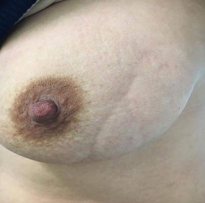
Figure 1: 35-year-old woman with a cord-like structure in the outer quadrant of the left breast which became more prominent when the patient raised her arm.
Results
The patient was diagnosed with idiopathic Mondor’s disease of the breast. Management was conservative, consisting of a short course of non-steroidal anti-inflammatory drugs (NSAIDs) and warm compresses for symptomatic relief.
The clinical progression was favorable: Within three days the patient reported complete resolution of the breast pain. On the follow-up examination, after four weeks, the palpable cord-like structure had entirely subsided.
No further interventions were required, and the patient was reassured of the benign nature of the condition.
Discussion
Mondor’s disease is a self-limiting condition, yet it remains a source of significant psychological distress for patients due to the sudden appearance of a palpable mass. The pathophysiology is incompletely understood and involves an initial inflammatory process of the vein wall followed by the formation of a thrombus and subsequent fibrosis, leading to the characteristic "iron wire" feel on palpation. The process of re-cannulization can take between four and eight weeks, with most patients having a complete resolution of symptoms [3].
In this case, the patient lacked traditional risk factors such as trauma or surgery, classifying the presentation as idiopathic. Although the association between Mondor’s disease and breast cancer is low (estimated at less than 5%), the literature emphasizes the necessity of mammography and ultrasound, especially in women over 35, to exclude occult malignancy that may be causing venous compression or hypercoagulability [3,4].
Treatment is focused on symptom management, as the condition typically resolves spontaneously within 4 to 8 weeks. The follow-up is usually uneventful with low rates of recurrence and of subsequent cancer [5].
Conclusion
This case highlights the importance of clinical awareness of Mondor’s disease in the differential diagnosis of breast pain. A thorough physical examination and appropriate imaging are essential to provide reassurance and avoid unnecessary invasive procedures. Once malignancy is excluded, conservative management with anti-inflammatories leads to excellent clinical outcomes.
Acknowledgment
The authors would like to thank the clinical staff of the Breast Clinic for their assistance in patient care.
Ethical Consideration
Written informed consent was obtained from the patient for the publication of this case report.
Conflict of Interest
The authors declare that they have no conflicts of interest that may influence the results or interpretations of the manuscript.
References
- Faage CH. (1869). Remarks on certain cutaneous affections. Guys Hosp Rep (3rd series); 15: 295-302.
- Mondor H. (1939). Tronculite sous-cutanée subaigue de la paroi thoracique antero-laterale. Mem Acad Chir. 65: 1271-1278.
- Amano M, Shimizu T. (2018). Mondor's Disease: A Review of the Literature. Intern Med. 57: 2607-2612.
- Markopoulos C, Kouskos E. (2005). Mondor's disease of the breast: is there any relation to breast cancer? Eur J Gynaec Oncol. 26: 213-214.
- Laroche JP, Galanaud JP, Labau D. (2012). Mondor's disease: what's new since 1939? Thromb Res. 130: S56-8.
© by the Authors & Gavin Publishers. This is an Open Access Journal Article Published Under Attribution-Share Alike CC BY-SA: Creative Commons Attribution-Share Alike 4.0 International License. Read More About Open Access Policy.