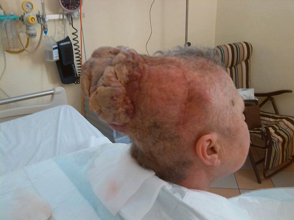
Exophytic Transcranial Relapse and Progression of Glioblastoma
Elena Orlandi1,2, Massimo Ambroggi1,2, Luigi Cavanna1,2*
1Department of Oncology and Hematology, Oncology Unit, Azienda, Italy
2Ospedaliera ‘Guglielmo da Saliceto’, Via Taverna 49 29121 Piacenza, Italy
*Corresponding author: Luigi Cavanna, Department of Oncology and Hematology, Oncology Unit, Azienda and Ospedaliera ‘Guglielmo da Saliceto’, Via Taverna 49 29121 Piacenza, Italy
Received Date: 22 September 2022
Accepted Date: 24 September 2022
Published Date: 27 September 2022
Citation: Orlandi E, Ambroggi M, Cavanna L (2022) Exophytic Transcranial Relapse and Progression of Glioblastoma. Ann Case Report. 7: 964. DOI: https://doi.org/10.29011/2574-7754.100964
Introduction
A 62-year-old woman with an exophytic transcranial relapse of glioblastoma and liquorrea is described. In April 2010 she underwent neurosurgery for glioblastoma G4, sub totally removed. The patient received Radiotherapy (RT) plus Chemotherapy (CT). In April 2012, local relapse was surgically removed, followed by 6 courses of CT. In January 2014, the patient underwent surgery again for local relapse, followed by RT and CT.
In June 2014, the patient presented a cranial mass close to the transcranial relapse, associated with Cerebrospinal Fluid (CSF) raising: MRI revealed relapse in the parietal and occipital region, extended from the brain across the cranial bone to the extra cranial superficial space (Figure); surgery, RT and CT were not indicated and the patient underwent palliative care in hospice.

What is the most likely working diagnosis?
- Pseudocystis consequently surgery and radiotherapy
- Glioblastoma esophytic transcranial relapse
- Extradural haematoma
- Transcranial benign tumor
Answer: B. Glioblastoma esophytic transcranial relapse
Discussion
The working diagnosis of Glioblastoma esophytic transcranial relapse is made based on the clinical and radiological findings and the history. The clinical features of the esophytic mass were suggestive of malignant neoplasm: the patient presented an eterogeneus mass with invasion and destruption of the surrounding bone associated with liquorrea; relapse can be associated with Cerebrospinal Fluid (CSF) raising. The MRI revealed relapse in the parietal and occipital region, extended from the brain across the cranial bone to the extra cranial superficial space.
Local dissemination to the scalp is rare because of the dura; usually can occur after previous surgical disruption of the dura and calvarium [1], which is supposed to cause seeding of the extra cranial space with tumor cell [2].
Surgery, Radiotherapy and Chemotherapy were not indicated and the patient underwent palliative care in hospice.
Teaching (take home) points: 1. Relapse extended from the brain across the cranial bone to the extra cranial superficial space; 2. Relapse was associated with Cerebrospinal Fluid (CSF) raising.
We obtained informed consent and permission from the patient to publish her image.
Disclosure of Potential Conflicts of Interest: We have no conflict of interest to declare.
References
- Rainov NG, Holzhausen HJ, Meyer H, Burkert W (1996) Local invasivity of glioblastoma multiforme with destruction of skull bone. Case report and review of the literature. Neurosurg Rev. 19: 183-8.
- Forsyth TM, Bi WL, Abedalthagafi M, Dunn IF, Chiocca EA (2015) Extracranial growth of glioblastoma multiforme. J Clin Neurosci. 22: 1521-1523.
© by the Authors & Gavin Publishers. This is an Open Access Journal Article Published Under Attribution-Share Alike CC BY-SA: Creative Commons Attribution-Share Alike 4.0 International License. Read More About Open Access Policy.