77-Year-Old, Female with a Palpable Right Breast Mass
by Lindsey Shwayyat, Cherie M. Kuzmiak*
Department of Radiology, University of North Carolina, Chapel Hill, NC, USA
*Corresponding Author: Cherie M. Kuzmiak, Department of Radiology, University of North Carolina, Chapel Hill, NC, USA
Received Date: 17 March 2026
Accepted Date: 23 March 2026
Published Date: 25 March 2026
Citation: Shwayyat L, Kuzmiak CM (2026) 77-Year-Old, Female with a Palpable Right Breast Mass. J Surg 11: 11592 DOI: https://doi.org/10.29011/2575-9760.011592
History
A 77-year-old female presented with a palpable lump in the right breast that had been enlarging for a year. Physical examination of the right breast demonstrated an 8.0 cm, mobile mass in the lateral aspect. A diagnostic bilateral mammogram was performed (Figure 1). After the mammogram, the patient underwent a diagnostic right breast ultrasound (Figure 2).
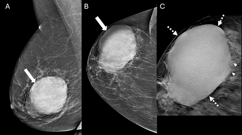
Figure 1: Mammogram images of a 77-year-old female with a palpable lump. Mediolateral oblique (A) and craniocaudal (B) mammogram images of the right breast demonstrate an 8.2 cm, oval, high-density, non-calcified mass (arrow) with predominantly circumscribed margins in the lateral aspect of the breast, anterior and middle depths. The circumscribed margins (dashed arrows) and subtle indistinct margins (arrowheads) are best appreciated on the spot tomosynthesis image (C). The left breast is normal (not shown).
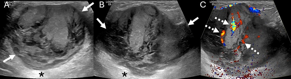
Figure 2: US images of a 77-year-old female with a palpable right breast mass in the lateral aspect of the right breast. Transverse (A) and longitudinal (B) US images of the right breast show an 8.0 cm, oval, parallel, heterogeneous mass (arrows) with indistinct margins and posterior acoustic enhancement (asterisk). Internal vascularity (dashed arrows) is present (C).
Imaging Findings
The mammogram showed an 8.2 cm, oval, high-density, non-calcified mass with predominantly circumscribed margins in the lateral aspect of the right breast (Figure 1). The left breast was normal. Targeted Ultrasound (US) of the right breast demonstrated an 8.0 cm, oval, parallel, heterogeneous mass with indistinct margins and posterior acoustic enhancement. Internal vascularity was present (Figures 2,3). The mass was assessed as a BI-RADS 4 lesion. US-guided biopsy of the mass was subsequently performed.
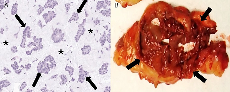
Figure 3: Histopathology image from a core needle biopsy of a 77-year-old female who presented with a right breast mass. (A) The mass yielded small tumor cells (arrows) in abundant extracellular mucin (asterisk), diagnostic for pure mucinous carcinoma (Hematoxylin And Eosin [H&E]; magnification, ×20) (B) The gross surgical specimen shows an 8.0 cm, oval, non-encapsulated, reddish soft tissue mass (arrows) with a gelatinous cut surface.
Differential Diagnosis
The differential diagnosis for an oval, non-calcified mass in a female patient includes invasive ductal carcinoma, fibroadenoma, and in the appropriate clinical setting, trauma/hematoma and infection [1]. Less common etiologies to consider are papilloma, papillary carcinoma, mucinous carcinoma, phyllodes, medullary carcinoma and metaplastic carcinoma [1-4]. High-grade invasive ductal and medullary carcinomas can present as an oval mass with circumscribed or not circumscribed margins [3]. Clinical history might assist in the diagnosis since medullary cancer is most often diagnosed in women age 40-50 with a BRCA1 mutation [3]. Both mucinous and papillary cancers tend to occur in postmenopausal women [1,2,5]. Mucinous cancers tend to be slow growing while phyllodes and metaplastic cancers rapidly enlarge [1,4].
Diagnosis
Pure mucinous carcinoma
Discussion
Mucinous Carcinoma (MC) is a subtype of invasive breast cancer (IBC) and accounts for <4% of breast cancers [1,2,6]. It occurs most commonly in postmenopausal women [1,2,6]. MC is characterized by pools of mucin around neoplastic cells. Based on mucin content, it is classified as pure (>90% mucin) or mixed (>10% to <90% mucin) [6]. The non-mucinous component of Mixed MC (MMC) is typically IBC of no special type (IBC-NST) [1,2,6,7]. Pure MCs (PMCs) tend to be of low or intermediate grade, and the majority are Hormone Receptor (HR) positive and negative for Human Epidermal Growth Factor Receptor-2 (HER2) [luminal A subtype] [6]. PMC is typically slow growing with a more favorable prognosis than MMC, with low rate of nodal involvement [1,2,6,7].
On mammography, PMC typically presents as an oval/round, equal to high-density mass with circumscribed or microlobulated margins [1,2,7]. Spiculated or indistinct margins are more indicative of MMC secondary to the IBC-NST component [1,2,7]. The ultrasound appearance of PMC is often as an oval/round mass with microlobulated or indistinct margins with variable echogenicity, ranging from isoechoic to mixed solid and cystic [1,2,7]. Posterior acoustic enhancement is often present due to high mucin content [1,2,7]. MRI features of PMC are of an oval/ round T1-hypointense, T2-hyperintense (due to mucin) mass often with rim or heterogenous enhancement [1,2,7,8]. Due to favorable histology, PMC is treated by surgical excision without nodal sampling [1]. Adjuvant endocrine therapy is recommended to decrease the risk of local recurrence and distant metastasis [1]. PMC has a low recurrence rate of 6.2% [1].
References
- Kuzmiak CM, Calhoun BC (2023) Pure mucinous carcinoma of the breast: radiologic-pathologic correlation. J Breast Imaging 5: 180-187.
- Thai JN, Lerwill MF, Chou SS (2023) Spectrum of mucin-containing lesions of the breast: multimodality imaging review with pathologic correlation. Radiographics 43: e230015.
- Pintican R, Duma M, Chiorean A (2020) Mucinous versus medullary breast carcinoma: mammography, ultrasound, and MRI findings. Clin Radiol 75: 483-496.
- Thapa B, Arobelidze S, Clark BA (2022) Metaplastic breast cancer: characteristics and survival outcomes. Cureus 14: e28551.
- Pal SK, Lau SK, Kruper L (2010) Papillary carcinoma of the breast: an overview. Breast Cancer Res Treat 122: 637-645.
- Wen HY, Desmedt C, Reis-Filho J, Schmitt F (2019) Epithelial tumours of the breast: mucinous carcinoma. In: WHO Classification of Tumours: Breast Tumours. 5th ed. Lyon, France: International Agency for Research on Cancer 123-127.
- Chaudhry AR, El Khoury M, Gotra A, et al (2019) Imaging features of pure and mixed forms of mucinous breast carcinoma with histopathological correlation. Br J Radiol 92 (1095).
- Bitencourt AG, Graziano L, Osorio CA (2016) MRI features of mucinous cancer of the breast: correlation with pathologic findings and other imaging methods 206: 238-246.
© by the Authors & Gavin Publishers. This is an Open Access Journal Article Published Under Attribution-Share Alike CC BY-SA: Creative Commons Attribution-Share Alike 4.0 International License. Read More About Open Access Policy.